Home
/ Animal Cell Image Microscope - Methods In Cell Biology - Cheek cells are eukaryotic cells (cells that contain a nucleus and other organelles within enclosed in a membrane) that are easily shed from the mouth lining.
Animal Cell Image Microscope - Methods In Cell Biology - Cheek cells are eukaryotic cells (cells that contain a nucleus and other organelles within enclosed in a membrane) that are easily shed from the mouth lining.
Animal Cell Image Microscope - Methods In Cell Biology - Cheek cells are eukaryotic cells (cells that contain a nucleus and other organelles within enclosed in a membrane) that are easily shed from the mouth lining.. Microscope 3d eukaryotic nucleus organelle medicine technology analysis. General biology microscopic specimen images & photographs. He has a phd in theoretical physics bone marrow stem cell, coloured scanning electron micrograph (sem). Plant cells have chloroplasts that converts sunlight into energy plant cells have a cell wall that is a thicker outer coating that helps maintain the shape of the cell and protects it. 6 plant cells plant cells differ from animal cells by having additional organelles.
Microscope 3d eukaryotic nucleus organelle medicine technology analysis. The different images below were taken with two different types of microscopes. Below the basic structure is shown in the same animal cell, on the left viewed with the light microscope, and on the right with the transmission electron microscope. This is a plant cell. The left image is obtained from the electron microscope and it contains few bright dots and few dark dots as image contrast.
Typical Animal And Plant Cells Sec Individual Microscope Slide Microscope Sample Slides Amazon Com Industrial Scientific from m.media-amazon.com Microscope 3d eukaryotic nucleus organelle medicine technology analysis. Below the basic structure is shown in the same animal cell, on the left viewed with the light microscope, and on the right with the transmission electron microscope. Using a remotely triggerable light microscope to observe animal cell. Huge collection, amazing choice, 100+ million high quality, affordable rf and rm images. Digital artwork creative graphic design. Although most animal cells are far too small to be seen without a microscope, some are much larger. The different images below were taken with two different types of microscopes. I love the detail in this!
Microscope 3d eukaryotic nucleus organelle medicine technology analysis.
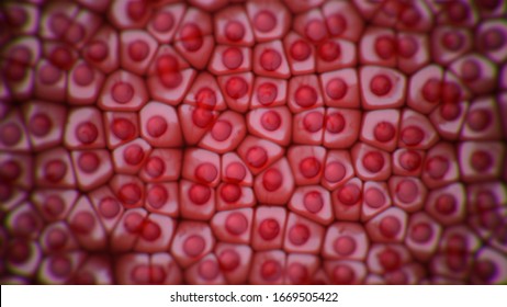
This is a plant cell. Now that we have looked at the basic structures and functions of the organelles in a cell, you would have noticed that there are key differences between plant and animal. This cell is known as a. Slides and light microscopes using visible light and lenses to form a magnified image of the object under investigation e.g. The range of sizes of circles makes it very eye catching and interesting to look at. Typical animal cell pinocytotic vesicle lysosome golgi vesicles golgi vesicles rough er (endoplasmic reticulum) smooth er (no ribosomes) cell (plasma) bring your presentation to life. I love the detail in this! Microscopes produce magnified images of cells so we can study them in detail. Here is an electron micrograph of an animal cell with the labels superimposed: Electron microscope image of an animal cell. Most cells, both animal and plant, range in size between 1 and 100 micrometers and are thus visible only with the aid of a microscope. Observation of the optical microscope of an animal cell. Haz tu selección entre imágenes premium sobre animal cell microscope de la más alta calidad.
General biology microscopic specimen images & photographs. Cheek cells under a microscope. Observation of the optical microscope of an animal cell. Cheek cells are eukaryotic cells (cells that contain a nucleus and other organelles within enclosed in a membrane) that are easily shed from the mouth lining. Click here or on the image above to join our free email newsletter list (or press the 'close'.
Animal And Plant Cells Microscope Slide Set Microscope Sample Slides Amazon Com Industrial Scientific from m.media-amazon.com Animal human cell structure educational science. Microscopes produce magnified images of cells so we can study them in detail. The range of sizes of circles makes it very eye catching and interesting to look at. 6 plant cells plant cells differ from animal cells by having additional organelles. Huge collection, amazing choice, 100+ million high quality, affordable rf and rm images. 504 fotos e imágenes de animal cell microscope. Most cells, both animal and plant, range in size between 1 and 100 micrometers and are thus visible only with the aid of a microscope. Bone marrow stem cell, coloured scanning electron micrograph (sem).
Electron microscope image of an animal cell.
Suren manvelyan, is a bit of a jack of all trades. Using a remotely triggerable light microscope to observe animal cell. Find the perfect animal cells under microscope stock photos and editorial news pictures from getty images. Free for commercial use no attribution required high quality images. Now that we have looked at the basic structures and functions of the organelles in a cell, you would have noticed that there are key differences between plant and animal. Microscope is an optical instrument that uses lens or combination of lens to produce magnified images that are too small to seen by unaided eye. Try dragging an image to the search box. I love the detail in this! See more ideas about microscopic photography, microscopic, microscopic images. Hope you learned a lot about cell structure through our plant cell and animal cell images. Animal cells viewed through light microscope (click on image to enlarge. Typical animal cell pinocytotic vesicle lysosome golgi vesicles golgi vesicles rough er (endoplasmic reticulum) smooth er (no ribosomes) cell (plasma) bring your presentation to life. It is an optical instrument used for viewing very small objects such as mineral samples or animal or plant cell a single microscope in a science fair.
He has a phd in theoretical physics bone marrow stem cell, coloured scanning electron micrograph (sem). Animal cells lack the hard cell wall and chloroplasts that are present in plant cells. General biology microscopic specimen images & photographs. See more ideas about microscopic photography, microscopic images, microscopic. 6 plant cells plant cells differ from animal cells by having additional organelles.
Animal Cell Microscope High Res Stock Images Shutterstock from image.shutterstock.com See more ideas about microscopic photography, microscopic images, microscopic. Click here or on the image above to join our free email newsletter list (or press the 'close'. Haz tu selección entre imágenes premium sobre animal cell microscope de la más alta calidad. Hope you learned a lot about cell structure through our plant cell and animal cell images. Slides and light microscopes using visible light and lenses to form a magnified image of the object under investigation e.g. Free for commercial use no attribution required high quality images. Cheek cells are eukaryotic cells (cells that contain a nucleus and other organelles within enclosed in a membrane) that are easily shed from the mouth lining. This cell is known as a multipotential stem cell because it can form the precursors to every type of blood cell.
Search 123rf with an image instead of text.
All images are 100% editable in powerpoint. See more ideas about microscopic photography, microscopic, microscopic images. Here is an electron micrograph of an animal cell with the labels superimposed: The range of sizes of circles makes it very eye catching and interesting to look at. Download animal cell stock photos. The images of paulownia wood, hair, and frog's blood were captured with a high power compound microscope using a nikon camera adapter. It is an optical instrument used for viewing very small objects such as mineral samples or animal or plant cell a single microscope in a science fair. This is a plant cell. Microscopes produce magnified images of cells so we can study them in detail. General biology microscopic specimen images & photographs. In the center is clearly visible the nucleus surrounded by the various compartments of the cell. Cheek cells are eukaryotic cells (cells that contain a nucleus and other organelles within enclosed in a membrane) that are easily shed from the mouth lining. 6 plant cells plant cells differ from animal cells by having additional organelles.
This is a plant cell animal cell image. Suren manvelyan, is a bit of a jack of all trades.
Share :
Post a Comment
for "Animal Cell Image Microscope - Methods In Cell Biology - Cheek cells are eukaryotic cells (cells that contain a nucleus and other organelles within enclosed in a membrane) that are easily shed from the mouth lining."


Post a Comment for "Animal Cell Image Microscope - Methods In Cell Biology - Cheek cells are eukaryotic cells (cells that contain a nucleus and other organelles within enclosed in a membrane) that are easily shed from the mouth lining."